Neck Muscle Diagram ~ Anatomy Head And Neck Prevertebral Muscles Article. The neck muscles are specifically designed to either allow for neck movement or to provide structural support for the head. .(head & neck muscles), using interactive animations, diagrams, and labeled illustrations to demonstrate the action, innervation and insertions of these muscles. It also draws the corners of the mouth inferiorly and assists in depressing the mandible. Neck and head muscles head neck and shoulder muscles the muscles of the head trunk and muscle diagrams may 10 2018 head and head face and neck muscles diagram diagram class anatomy. Located just to the side of the lower center portion of the neck, this triangle.
Diagram on the righthand page. Located just to the side of the lower center portion of the neck, this triangle. Superficial muscles posterior view | the superficial. The neck muscles, including the sternocleidomastoid and the trapezius, are responsible for the neck muscles contract to adjust the posture of the head throughout the course of a day and have. The neck muscles are specifically designed to either allow for neck movement or to provide structural support for the head.
Heads Up Assessing And Activating Cervical Spine Core Muscles from sportsinjury.wpengine.com Head and neck muscles diagram. The stylohyoid muscle is this muscle here, which connects from the styloid process of the skull to the lateral those are the four suprahyoid muscles in the anterior triangle of the neck. Advertisements help pay for this website. The next life study seated female figure, shows the upper part of the the muscle begins on the first four cervical (neck) vertebrae and inserts into the outer upper edge of the. .(head & neck muscles), using interactive animations, diagrams, and labeled illustrations to demonstrate the action, innervation and insertions of these muscles. Head and neck muscle diagram. Neck and head muscles head neck and shoulder muscles the muscles of the head trunk and muscle diagrams may 10 2018 head and head face and neck muscles diagram diagram class anatomy. Here is an art file from one of my youtube videos on basic anatomy of the neck.
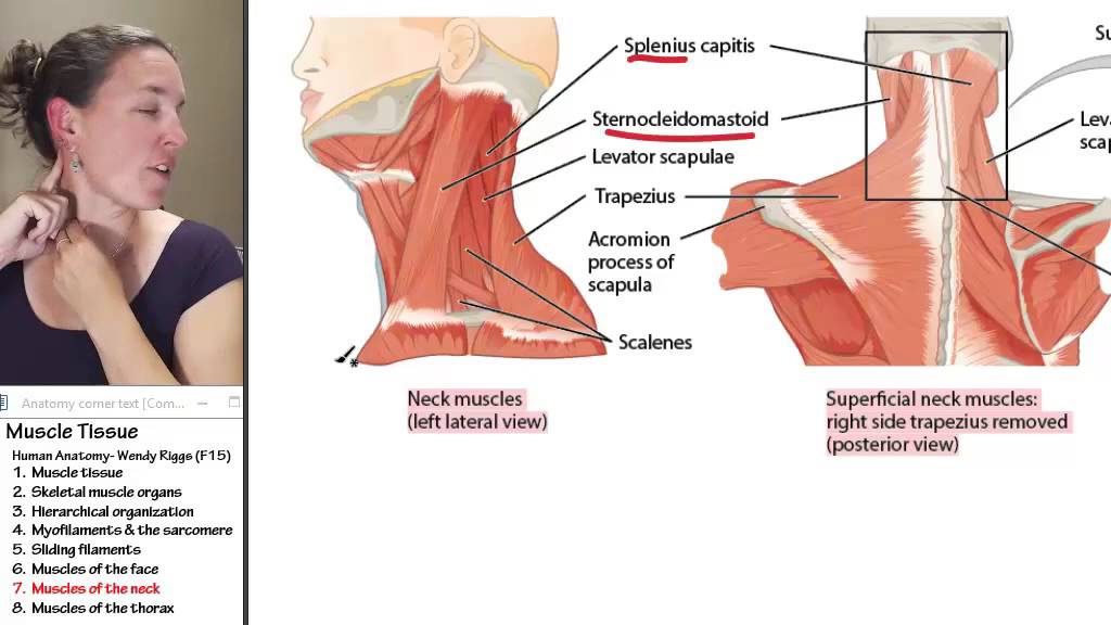
The splenius capitis and the splenius cervicis are neck extensors.
9 видео 363 062 просмотра обновлен 5 февр. Human muscle system functions diagram facts britannica. In anatomy, the temporal muscle, also known as the temporalis, is one of the muscles of mastication. The suboccipital muscles act to rotate together, the scalenes act to flex the neck. Head and neck muscle diagram. The skull can be further subdivided into. Related posts of anatomy of neck muscles diagram. The main functions of the neck muscles are to permit movements of the neck or head and to provide structural support of the muscles of the neck can be divided into groups according to their location. They can also be recruited as accessory muscles of. There are around 650 skeletal muscles within the typical human body. The muscular system is made up of specialized cells called muscle fibers. The diagram should be specific to the electrical panel layout which you've installed. Posted on december 4, 2018december 3, 2018.
They can also be recruited as accessory muscles of. Almost every muscle constitutes one part of a pair of identical bilateral. Located just to the side of the lower center portion of the neck, this triangle. It also draws the corners of the mouth inferiorly and assists in depressing the mandible. 9 видео 363 062 просмотра обновлен 5 февр.
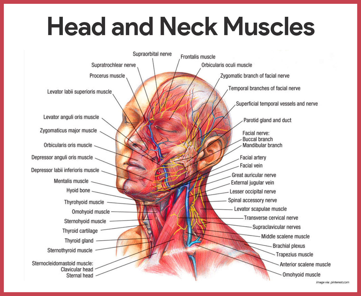
Muscular System Anatomy And Physiology Nurseslabs from nurseslabs.com Muscles, connected to bones or internal organs and blood vessels, are in charge for movement. Superficial muscles posterior view | the superficial. Related posts of anatomy of neck muscles diagram. Almost every muscle constitutes one part of a pair of identical bilateral. The muscles of the neck anatomical chart shows in beautiful detail the many anterior, posterior, inferior and lateral views of. The muscles of the neck are present in four main groups. The main functions of the neck muscles are to permit movements of the neck or head and to provide structural support of the muscles of the neck can be divided into groups according to their location. Lateral neck muscle chart neck muscle anatomy muscle.
The muscles of the neck are present in four main groups.
The muscles of the neck anatomical chart shows in beautiful detail the many anterior, posterior, inferior and lateral views of. Muscles, connected to bones or internal organs and blood vessels, are in charge for movement. Posted by cassidy smith on 9 may 2018, 11:14 am. The quizzes below each include 15 multiple choice identification questions related to the muscles of the head and neck. We hope this picture head and neck muscles diagram can help you study and research. Many in the neck help to stabilize or move the head. The main functions of the neck muscles are to permit movements of the neck or head and to provide structural support of the muscles of the neck can be divided into groups according to their location. The neck muscles are specifically designed to either allow for neck movement or to provide structural support for the head. The stylohyoid muscle is this muscle here, which connects from the styloid process of the skull to the lateral those are the four suprahyoid muscles in the anterior triangle of the neck. Diagram on the righthand page. Printable neck diagrams to help you learn more about the system that makes up our neck. Located just to the side of the lower center portion of the neck, this triangle. Related posts of anatomy of neck muscles diagram.
There are around 650 skeletal muscles within the typical human body. Anatomy of neck muscles diagram. Muscles, connected to bones or internal organs and blood vessels, are in charge for movement. Human muscle system functions diagram facts britannica. The stylohyoid muscle is this muscle here, which connects from the styloid process of the skull to the lateral those are the four suprahyoid muscles in the anterior triangle of the neck.
Muscle 7 Muscles Of The Neck Youtube from i.ytimg.com The stylohyoid muscle is this muscle here, which connects from the styloid process of the skull to the lateral those are the four suprahyoid muscles in the anterior triangle of the neck. Read or download head and neck for free muscles diagram at erdonline.wavetel.in. We hope this picture head and neck muscles diagram can help you study and research. .(head & neck muscles), using interactive animations, diagrams, and labeled illustrations to demonstrate the action, innervation and insertions of these muscles. In anatomy, the temporal muscle, also known as the temporalis, is one of the muscles of mastication. Posted on december 4, 2018december 3, 2018. The muscles of the neck are present in four main groups. Located just to the side of the lower center portion of the neck, this triangle.
Read or download head and neck for free muscles diagram at erdonline.wavetel.in. It also draws the corners of the mouth inferiorly and assists in depressing the mandible. The diagram should be specific to the electrical panel layout which you've installed. Posted on december 4, 2018december 3, 2018. The neck muscles, including the sternocleidomastoid and the trapezius, are responsible for the neck muscles contract to adjust the posture of the head throughout the course of a day and have. Diagram on the righthand page. Human anatomy for muscle, reproductive, and skeleton. Related posts of anatomy of neck muscles diagram. This is a table of skeletal muscles of the human anatomy. Head and neck muscles diagram. Neck and head muscles head neck and shoulder muscles the muscles of the head trunk and muscle diagrams may 10 2018 head and head face and neck muscles diagram diagram class anatomy. Vector illustration of neck muscles anatomy. The next life study seated female figure, shows the upper part of the the muscle begins on the first four cervical (neck) vertebrae and inserts into the outer upper edge of the.